Overpronation Of The Feet
Overview
Pes planus is the medical term for flat feet. It comes from Latin, Pes = foot and Planus = plain, level ground. Very few people suffer from this condition, as a true flat foot is very rare. Less than 5% of the population has flat feet. The majority of the population, however, has fallen arches (an estimated 60-70% of the population) known in the medical profession as ?excess pronation? or over-pronation. Over-pronation means the foot and ankle tend to roll inwards and the arch collapses with weight-bearing. This is a quite a destructive position for the foot to function in and may cause a wide variety of foot, leg and lower back conditions.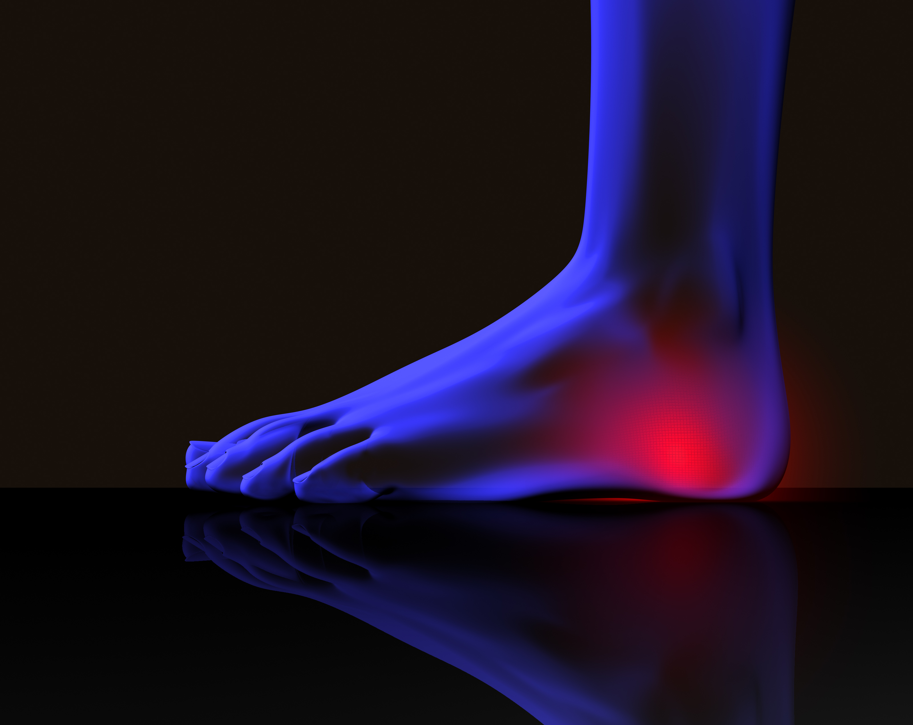
Causes
Congenital "Flat Feet" - an individual may be born with feet that lack an appropriately supportive arch thereby predisposing the individual to this foot condition. Excessive Weight (Obesity) Too much weight on the foot from either obesity or pregnancy may be a factor. Repetitive Impact walking on flat, hard surfaces continuously places unnatural stress on the foot arch.
Symptoms
In addition to problems overpronation causes in the feet, it can also create issues in the calf muscles and lower legs. The calf muscles, which attach to the heel via the Achilles tendon, can become twisted and irritated as a result of the heel rolling excessively toward the midline of the body. Over time this can lead to inflexibility of the calf muscles and the Achilles tendon, which will likely lead to another common problem in the foot and ankle complex, the inability to dorsiflex. As such, overpronation is intrinsically linked to the inability to dorsiflex.
Diagnosis
If you have flat feet or low arches, chances are you overpronate. Although not always the case, the lower your arches the greater the overpronate. Stand on a hard surface (in front of a mirror if you need to) and look at your feet, flat feet or low arches are easy to spot. If your feet look flatter than a pancake, have a look at your ankles and see if they seem collapsed or straight. If they are, you're overpronating.
Non Surgical Treatment
Side Step with Opposite Reach. This exercise is designed to load the "bungee cord system" of the gluteal muscle and its opposite, latissimus dorsi muscle to keep the foot from overpronating. Because the opposite arm swings across the front leg when walking, this exercise creates tension in the muscles all the way from the front foot, across the back of the hips and back, to the fingers of the opposite hand. Movement Directions. Stand with left foot on top of the dome of the BT. (Note: For added balance, the right foot can tap on the ground, if needed). Reach right leg out to the side of the BT, and tap the ground while squatting down on the left side and reaching right arm across the left knee. Push down with left big toe while squatting. This activates the arch of the left foot and strengthens all the stabilizing muscles on the left side of the lower body. Return to starting position. Perform 8 to 10 repetitions on each leg.
Surgical Treatment
Depending on the severity of your condition, your surgeon may recommend one or more treatment options. Ultimately, however, it's YOUR decision as to which makes the most sense to you. There are many resources available online and elsewhere for you to research the various options and make an informed decision.
Physical Rehabilitation And Calcaneal Apophysitis
Overview
Sever?s disease or Sever?s lesion refers to an injury to the bone growth plate at the back of the heel bone (calcaneous) in young people, particularly those who are physically active. It usually develops in puberty and is slightly more common in boys than girls.
Causes
There is no specific known cause of Sever?s disease. However, there are several common factors associated with the condition including. Tight calf muscles. Pronated foot type (rolled in towards the ankle). Children who are heavier. Puberty/growth spurts. External factors, e.g. hard surfaces or poor footwear. Increase in physical activity levels.
Symptoms
Some of the most common signs and symptoms associated with Sever?s disease include. Heel pain or tenderness in one or both heels, usually at the back of the heel. Pain or discomfort upon waking, or when the heel is squeezed. Heel pain that is worse during or following activity. Limping. Heel swelling or redness. Tight calf muscles. Decreased ankle range of motion.
Diagnosis
A doctor can usually tell that a child has Sever's disease based on the symptoms reported. To confirm the diagnosis, the doctor will probably examine the heels and ask about the child's activity level and participation in sports. The doctor might also use the squeeze test, squeezing the back part of the heel from both sides at the same time to see if doing so causes pain. The doctor might also ask the child to stand on tiptoes to see if that position causes pain. Although imaging tests such as X-rays generally are not that helpful in diagnosing Sever's disease, some doctors order them to rule out other problems, such as fractures. Sever's disease cannot be seen on an X-ray.
Non Surgical Treatment
Depending on the Podiatrist's diagnosis and the severity of the pain, there are several treatment options available. Rest/ reduced activity: your child should reduce or stop any activity that causes pain, such as sports and running. This can be a difficult option, as children are normally quite willful in pursuit of their favorite pastimes! Over the counter anti-inflammatory drugs, such as ibuprofen (found in Nurofen), to help reduce pain and inflammation. Try to make sure your child does the recommended stretching exercises before sport/play. This will should help reduce the stress on the fascia tendon and relieve heel pain. The use of Orthotic insoles. Footactive Kids orthotics are made for children. They will help properly support the foot, help prevent over-pronation or improper gait restoring your child's foot the the correct biomechanical position. If you are in any doubt or your child's foot pain persists then please arrange an appointment with a Podiatrist or Physiotherapist. Please click here for more information on the use of orthotics for children.
Posterior Tibial Tendon Insufficiency Treatment
Overview
PTTD is a condition of degeneration and dysfunction in the tendon complex that helps control the medial arch of your foot. Essentially what happens is the complex is unable to do its job of supporting the arch and supinating the foot, so a progressive flat foot develops (usually called adult acquired flat foot). Initially pain and often swelling develops on the inside of the ankle and it will continue to get progressively worse. There are a number of stages of PTTD (3 Stages) and it needs to be aggressively treated early on otherwise a surgical reconstruction of the arch will invariably be required. PTTD can develop into a very disabling condition if it is not dealt with properly and promptly. As PTTD becomes more advanced, the arch flattens even more and the pain often shifts to the outside of the foot, below the ankle. Arthritis often develops in the foot and In more severe cases, arthritis may also develop in the ankle. 
Causes
Posterior tibial tendon dysfunction is the most common cause of acquired adult flatfoot. Sometimes this can be a result of specific trauma, but usually the tendon becomes injured from wear and tear over time. This is more prevalent in individuals with an inherited flat foot but excessive weight, age, and level of activity are also contributing factors.
Symptoms
In many cases, adult flatfoot causes no pain or problems. In others, pain may be severe. Many people experience aching pain in the heel and arch and swelling along the inner side of the foot.
Diagnosis
It is of great importance to have a full evaluation, by a foot and ankle specialist with expertise in addressing complex flatfoot deformities. No two flat feet are alike; therefore, "Universal" treatment plans do not exist for the Adult Flatfoot. It is important to have a custom treatment plan that is tailored to your specific foot. That starts by first understanding all the intricacies of your foot, through an extensive evaluation. X-rays of the foot and ankle are standard, and MRI may be used to better assess the quality of the PT Tendon.
Non surgical Treatment
Non-surgical treatment consists of custom orthoses and or special bracing devices along with supportive measures aimed at reducing the symptoms. While non-surgical treatment helps the majority of patients with PTTD, progressive cases may require surgical treatment including soft tissue tendon transfers, osteotomies and lastly fusion. 
Surgical Treatment
For more chronic flatfoot pain, surgical intervention may be the best option. Barring other serious medical ailments, surgery is a good alternative for patients with a serious problem. There are two surgical options depending on a person?s physical condition, age and lifestyle. The first type of surgery involves repair of the PTT by transferring of a nearby tendon to help re-establish an arch and straighten out the foot. After this surgery, patients wear a non-weight bearing support boot for four to six weeks. The other surgery involves fusing of two or three bones in the hind foot below the ankle. While providing significant pain relief, this option does take away some hind foot side-to-side motion. Following surgery, patients are in a cast for three months. Surgery is an effective treatment to address adult-acquired flatfoot, but it can sometimes be avoided if foot issues are resolved early. That is why it is so important to seek help right away if you are feeling ankle pain. But perhaps the best way to keep from becoming flatfooted is to avoid the risk factors altogether. This means keeping your blood pressure, weight and diabetes in check.
Sharp Pain In Right Foot Arch
Overview
Painful progressive flatfoot, otherwise known as Tibialis Posterior Tendinitis, is caused by inflammation of the tendon of the tibialis posterior. The tendon then becomes inflamed, stretched or suffers a partial or total tear. If left untreated, this condition may lead to severe disability and chronic pain. Some people are predisposed to this condition if they have flatfeet or an abnormal attachment of the tendon to the bones in the midfoot. Nonsteroidal anti-inflammatory medications, ice, physical therapy, supportive taping and bracing, or orthotic devices are the common ways of treating painful progressive flatfoot. Contact your physician before taking any medication. In some cases, a surgical operation may need to be performed to repair the torn or damaged tendon and restore normal function. To prevent reinjury, orthotic devices may be recommended. In severe cases, surgery on the midfoot bones may be necessary to treat the associated flatfoot condition.

Causes
Plantar fasciitis, another sports injury detailed on this website, is regularly the cause of foot arch pain or strain. This can arise due to faulty biomechanics in your feet, which alone can also provoke foot arch pains. The most prominent biomechanical difficulties are flat feet and high arches. With flat feet (or overpronation) the arches appear to be almost flattened, causing unevenness by forcing the feet roll inwards in order to maintain balance and support the body's weight. This places inordinate pressure on the plantar fascia and arches. If by contrast you have high arches (instep), the ankle can roll outwards, again causing undue strain on the arches. Too much of this strain can lead to stretching of the plantar fascia and pain in the arches. Other causes include overstretching or otherwise pressuring the arches, for example by exercising with fatigued leg muscles which leave the feet with excessive work to do. You are also particularly at risk if in your 40s or 50s and commencing an intense program of training after a long period of inactivity.
Symptoms
Repetitive exertive activity arch pain is usually sharp, and localized to a specific area, rather than the entire arch. Usually the pain occurs in the area just in front of the heel. It is present when first standing on the foot in the morning, but may decrease once you start walking around, but will, gradually becomes worse with continued walking or running. Swelling may be present. The pain subsides with rest, but stretching the arch while resting may cause the pain to return. Injury pain is constantly present, but worse when standing on the foot. This pain is localized to a specific area, but may radiate out from this area to the entire foot. The pain is sharp, and usually accompanied by swelling and occasionally "black and blue" discolorations. The pain due to the natural aging process is usually dull and aching, or stiff, and can be felt throughout the entire arch area, rather then in just one spot. This pain is present whenever weight bearing, and usually becomes worse with continued walking. The pain gradually subsides when resting, and usually does not return with stretching. Biomechanical defect pain is usually localized to a section of the arch, such as the inner, middle, outer, front, or back of the arch. This pain may be sharp or dull, but is always worse with continued walking.
Diagnosis
The adult acquired flatfoot, secondary to posterior tibial tendon dysfunction, is diagnosed in a number of ways with no single test proven to be totally reliable. The most accurate diagnosis is made by a skilled clinician utilizing observation and hands on evaluation of the foot and ankle. Observation of the foot in a walking examination is most reliable. The affected foot appears more pronated and deformed compared to the unaffected foot. Muscle testing will show a strength deficit. An easy test to perform in the office is the single foot raise.
Non Surgical Treatment
How the pain in the bottom of your foot is treated will depend heavily on the cause of the pain. Diagnosing the pain while it?s in the early stages is important when determining the best treatment options. If the pain is mild to moderate, simple improvements in footwear can help reduce the symptoms. Most patients must use the RICE method for effective treatment. RICE stands for Rest, Ice, Compression, and Elevation. This is a popular treatment used by athletes. It involves resting the foot, icing it for fifteen to twenty minute intervals, compressing the foot with a bandage, and elevating it at least twelve inches above the heart. Ant-inflammatory and pain medications are also sometimes used to treat bottom-of-foot pain. For more serious cases, steroid injections or foot surgery may help reduce pain and swelling and correct the underlying condition (if there is one.) If you suffer from a severe case of plantar fasciitis and non-surgical methods fail, your doctor may recommend cortisone injections to relieve the pain. If cortisone injections fail, your doctor may recommend a surgical procedure that involves cutting and releasing the plantar fascia.

Surgical Treatment
Cavus foot is caused in part by an over-pull of one of the lateral ankle muscles. A release of this tendon can be performed on the outside of the ankle. Additionally, a transfer of this tendon can be performed to help in correcting deformity of the ankle joint. Often patients will have a tightness of their gastrocnemius muscle, one of the main muscles in the calf. This can increase the deformity or prevent a correction from working. It is addressed with a lengthening of a part of the calf muscle or Achilles tendon. This is often performed through one or more small cuts in the back of the leg or ankle. Finally, the plantar fascia may be tight. The plantar fascia is a cord-like structure that runs from the heel to the front part of the foot. Partial or complete plantar fascia release may be done.
Prevention
Foot and ankle injuries are common in sports, especially running, tennis and soccer. But sports enthusiasts can decrease the risk of injury by taking some precautions. Lightly stretch or better yet, do a slow jog for two to three minutes to warm up the muscles. Don't force the stretch with a "bouncing motion." The amount of time spent on the activity should be increased gradually over a period of weeks to build both muscle strength and mobility. Cross training by participating in different activities can help build the muscles. People whose feet pronate or who have low arches should choose shoes that provide support in both the front of the shoe and under the arch. The heel and heel counter (back of the shoe) should be very stable. Those with a stiffer foot or high arches should choose shoes with more cushion and a softer platform. Use sport-specific shoes. Cross training shoes are an overall good choice; however, it is best to use shoes designed for the sport.
Stretching Exercises
Easy Beginner Version. Start with your bare foot on a flat surface, toes spread out. Place a penny under the ball of your foot and the end of a pen under the middle of your arch (sticking out from the inside of your foot). Activate your arch by flexing your arch muscle. You should feel the muscles on the ball of your foot pushing down on the penny, but your arch shouldn't be pushing down on the pen. These tools help you (1) avoid rolling your foot and (2) avoid pressing down with your toes (as an extra tip, you can slide a business card under your toes before doing the exercise-when you activate your arch, you should be able to slide the business card out easily with your fingers). Do your best to keep your toes relaxed. Advanced Version. Once you're ready to move on, you can try this advanced version. It builds on the above exercise to incorporate full body twisting and balance, helping you to maintain proper arches while you move. Using the same ideas from above, stand on a flat surface in your bare feet with a penny under the ball of your foot and the end of a pen under your arch. This time, stand with your back a few inches away form a wall or a door. Lift your other leg (the one without the penny or pen) and stand on one foot. Use the wall for balance, if necessary. Lift one arm and stretch it across your body until you touch the wall or door on the opposite side, maintaining a straight back. Keep your foot straight and your arch on the penny but above the pen. Your arch will want to follow the movement and roll off, but you will need to activate it to stay stable during the movement. Lift your other arm and stretch it across the opposite side of your body, still keeping your arch in place.
Painful progressive flatfoot, otherwise known as Tibialis Posterior Tendinitis, is caused by inflammation of the tendon of the tibialis posterior. The tendon then becomes inflamed, stretched or suffers a partial or total tear. If left untreated, this condition may lead to severe disability and chronic pain. Some people are predisposed to this condition if they have flatfeet or an abnormal attachment of the tendon to the bones in the midfoot. Nonsteroidal anti-inflammatory medications, ice, physical therapy, supportive taping and bracing, or orthotic devices are the common ways of treating painful progressive flatfoot. Contact your physician before taking any medication. In some cases, a surgical operation may need to be performed to repair the torn or damaged tendon and restore normal function. To prevent reinjury, orthotic devices may be recommended. In severe cases, surgery on the midfoot bones may be necessary to treat the associated flatfoot condition.
Causes
Plantar fasciitis, another sports injury detailed on this website, is regularly the cause of foot arch pain or strain. This can arise due to faulty biomechanics in your feet, which alone can also provoke foot arch pains. The most prominent biomechanical difficulties are flat feet and high arches. With flat feet (or overpronation) the arches appear to be almost flattened, causing unevenness by forcing the feet roll inwards in order to maintain balance and support the body's weight. This places inordinate pressure on the plantar fascia and arches. If by contrast you have high arches (instep), the ankle can roll outwards, again causing undue strain on the arches. Too much of this strain can lead to stretching of the plantar fascia and pain in the arches. Other causes include overstretching or otherwise pressuring the arches, for example by exercising with fatigued leg muscles which leave the feet with excessive work to do. You are also particularly at risk if in your 40s or 50s and commencing an intense program of training after a long period of inactivity.
Symptoms
Repetitive exertive activity arch pain is usually sharp, and localized to a specific area, rather than the entire arch. Usually the pain occurs in the area just in front of the heel. It is present when first standing on the foot in the morning, but may decrease once you start walking around, but will, gradually becomes worse with continued walking or running. Swelling may be present. The pain subsides with rest, but stretching the arch while resting may cause the pain to return. Injury pain is constantly present, but worse when standing on the foot. This pain is localized to a specific area, but may radiate out from this area to the entire foot. The pain is sharp, and usually accompanied by swelling and occasionally "black and blue" discolorations. The pain due to the natural aging process is usually dull and aching, or stiff, and can be felt throughout the entire arch area, rather then in just one spot. This pain is present whenever weight bearing, and usually becomes worse with continued walking. The pain gradually subsides when resting, and usually does not return with stretching. Biomechanical defect pain is usually localized to a section of the arch, such as the inner, middle, outer, front, or back of the arch. This pain may be sharp or dull, but is always worse with continued walking.
Diagnosis
The adult acquired flatfoot, secondary to posterior tibial tendon dysfunction, is diagnosed in a number of ways with no single test proven to be totally reliable. The most accurate diagnosis is made by a skilled clinician utilizing observation and hands on evaluation of the foot and ankle. Observation of the foot in a walking examination is most reliable. The affected foot appears more pronated and deformed compared to the unaffected foot. Muscle testing will show a strength deficit. An easy test to perform in the office is the single foot raise.
Non Surgical Treatment
How the pain in the bottom of your foot is treated will depend heavily on the cause of the pain. Diagnosing the pain while it?s in the early stages is important when determining the best treatment options. If the pain is mild to moderate, simple improvements in footwear can help reduce the symptoms. Most patients must use the RICE method for effective treatment. RICE stands for Rest, Ice, Compression, and Elevation. This is a popular treatment used by athletes. It involves resting the foot, icing it for fifteen to twenty minute intervals, compressing the foot with a bandage, and elevating it at least twelve inches above the heart. Ant-inflammatory and pain medications are also sometimes used to treat bottom-of-foot pain. For more serious cases, steroid injections or foot surgery may help reduce pain and swelling and correct the underlying condition (if there is one.) If you suffer from a severe case of plantar fasciitis and non-surgical methods fail, your doctor may recommend cortisone injections to relieve the pain. If cortisone injections fail, your doctor may recommend a surgical procedure that involves cutting and releasing the plantar fascia.
Surgical Treatment
Cavus foot is caused in part by an over-pull of one of the lateral ankle muscles. A release of this tendon can be performed on the outside of the ankle. Additionally, a transfer of this tendon can be performed to help in correcting deformity of the ankle joint. Often patients will have a tightness of their gastrocnemius muscle, one of the main muscles in the calf. This can increase the deformity or prevent a correction from working. It is addressed with a lengthening of a part of the calf muscle or Achilles tendon. This is often performed through one or more small cuts in the back of the leg or ankle. Finally, the plantar fascia may be tight. The plantar fascia is a cord-like structure that runs from the heel to the front part of the foot. Partial or complete plantar fascia release may be done.
Prevention
Foot and ankle injuries are common in sports, especially running, tennis and soccer. But sports enthusiasts can decrease the risk of injury by taking some precautions. Lightly stretch or better yet, do a slow jog for two to three minutes to warm up the muscles. Don't force the stretch with a "bouncing motion." The amount of time spent on the activity should be increased gradually over a period of weeks to build both muscle strength and mobility. Cross training by participating in different activities can help build the muscles. People whose feet pronate or who have low arches should choose shoes that provide support in both the front of the shoe and under the arch. The heel and heel counter (back of the shoe) should be very stable. Those with a stiffer foot or high arches should choose shoes with more cushion and a softer platform. Use sport-specific shoes. Cross training shoes are an overall good choice; however, it is best to use shoes designed for the sport.
Stretching Exercises
Easy Beginner Version. Start with your bare foot on a flat surface, toes spread out. Place a penny under the ball of your foot and the end of a pen under the middle of your arch (sticking out from the inside of your foot). Activate your arch by flexing your arch muscle. You should feel the muscles on the ball of your foot pushing down on the penny, but your arch shouldn't be pushing down on the pen. These tools help you (1) avoid rolling your foot and (2) avoid pressing down with your toes (as an extra tip, you can slide a business card under your toes before doing the exercise-when you activate your arch, you should be able to slide the business card out easily with your fingers). Do your best to keep your toes relaxed. Advanced Version. Once you're ready to move on, you can try this advanced version. It builds on the above exercise to incorporate full body twisting and balance, helping you to maintain proper arches while you move. Using the same ideas from above, stand on a flat surface in your bare feet with a penny under the ball of your foot and the end of a pen under your arch. This time, stand with your back a few inches away form a wall or a door. Lift your other leg (the one without the penny or pen) and stand on one foot. Use the wall for balance, if necessary. Lift one arm and stretch it across your body until you touch the wall or door on the opposite side, maintaining a straight back. Keep your foot straight and your arch on the penny but above the pen. Your arch will want to follow the movement and roll off, but you will need to activate it to stay stable during the movement. Lift your other arm and stretch it across the opposite side of your body, still keeping your arch in place.
Ways To Address Fallen Arches
Overview

A normal foot should arch so the middle of the foot does not touch the ground when the patient stands up. If it does touch, the patient has a flat foot or fallen arch. Flat feet are normal for young children. Their arches should develop by adulthood, but sometimes they fail to develop. Some people with flat feet have pain in the heel or arch, but others do not.
Causes
When flat feet develop at a later age, they are known as fallen arches. The arches may fall because the muscles supporting them are no longer able to do so. In addition the spring ligament within the foot may have lost some of its tension allowing the arch of the foot to flatten. Other conditions causing fallen arches include sudden weight gain, a nervous system injury, or a loss of sensation caused by a disease such as diabetes. Most people with fallen arches are flat on both feet.
Symptoms
Flat feet may not cause any symptoms at all. Rigid flat feet may cause pain, calluses, blisters, or skin redness on the inner side of the foot. A stiff foot, weakness or numbness of the foot, Rapid wearing out of shoes-worn shoes lean in toward each other. Difficulty or pain with activities like running-in the foot, knee or hip.
Diagnosis
Diagnosis of flat feet or fallen arches can be made by your health practitioner and is based on the following. Clinical assessment involving visual gait assessment, as well as biomechanical assessment. A detailed family and medical history. A pain history assessment determining the location of painful symptoms. Physical palpation of the feet and painful areas. Imaging such as MRI or x-ray can be used by your practitioner to assist in the diagnosis.
Non Surgical Treatment
Treatment of flat feet by a fully educated, trained, and licensed podiatrist should be sought if the previously mentioned pain in the foot or knee areas start to surface and becomes a painful problem. Depending on the cause and exact type of the condition, a variety of forms of treatment may be prescribed. Verification of the exact cause and type of flat feet that each individual has, should only be handled by a professional podiatrist. Often times, the actual treatment method will include some form of arch support or light gymnastic style exercise. In rare cases, something more involved may be necessary, but only your skilled podiatrist would be able to accurately make that determination. If you suffer from flat feet, plantar fasciitis, or any other form of problematic foot condition, we encourage you to contact a foot clinic today and potentially rid yourself of that problem in virtually no time at all.
Surgical Treatment

This is rare and usually only offered if patients have significant abnormalities in their bones or muscles. Treatments include joint fusion, reshaping the bones in the foot, and occasionally moving around tendons in the foot to help balance out the stresses (called tendon transfer). Flat feet and fallen arches are common conditions that are in most cases asymptomatic. However, in patients who do have symptoms, treatments are available that can help reduce pain and promote efficient movement. Orthotic devices are well recognised as an excellent treatment and podiatrists can offer these different treatment modalities as individualised treatments for patients.
After Care
Time off work depends on the type of work as well as the surgical procedures performed. . A patient will be required to be non-weight bearing in a cast or splint and use crutches for four to twelve weeks. Usually a patient can return to work in one to two weeks if they are able to work while seated. If a person's job requires standing and walking, return to work may take several weeks. Complete recovery may take six months to a full year. Complications can occur as with all surgeries, but are minimized by strictly following your surgeon's post-operative instructions. The main complications include infection, bone that is slow to heal or does not heal, progression or reoccurrence of deformity, a stiff foot, and the need for further surgery. Many of the above complications can be avoided by only putting weight on the operative foot when allowed by your surgeon.
A normal foot should arch so the middle of the foot does not touch the ground when the patient stands up. If it does touch, the patient has a flat foot or fallen arch. Flat feet are normal for young children. Their arches should develop by adulthood, but sometimes they fail to develop. Some people with flat feet have pain in the heel or arch, but others do not.
Causes
When flat feet develop at a later age, they are known as fallen arches. The arches may fall because the muscles supporting them are no longer able to do so. In addition the spring ligament within the foot may have lost some of its tension allowing the arch of the foot to flatten. Other conditions causing fallen arches include sudden weight gain, a nervous system injury, or a loss of sensation caused by a disease such as diabetes. Most people with fallen arches are flat on both feet.
Symptoms
Flat feet may not cause any symptoms at all. Rigid flat feet may cause pain, calluses, blisters, or skin redness on the inner side of the foot. A stiff foot, weakness or numbness of the foot, Rapid wearing out of shoes-worn shoes lean in toward each other. Difficulty or pain with activities like running-in the foot, knee or hip.
Diagnosis
Diagnosis of flat feet or fallen arches can be made by your health practitioner and is based on the following. Clinical assessment involving visual gait assessment, as well as biomechanical assessment. A detailed family and medical history. A pain history assessment determining the location of painful symptoms. Physical palpation of the feet and painful areas. Imaging such as MRI or x-ray can be used by your practitioner to assist in the diagnosis.
Non Surgical Treatment
Treatment of flat feet by a fully educated, trained, and licensed podiatrist should be sought if the previously mentioned pain in the foot or knee areas start to surface and becomes a painful problem. Depending on the cause and exact type of the condition, a variety of forms of treatment may be prescribed. Verification of the exact cause and type of flat feet that each individual has, should only be handled by a professional podiatrist. Often times, the actual treatment method will include some form of arch support or light gymnastic style exercise. In rare cases, something more involved may be necessary, but only your skilled podiatrist would be able to accurately make that determination. If you suffer from flat feet, plantar fasciitis, or any other form of problematic foot condition, we encourage you to contact a foot clinic today and potentially rid yourself of that problem in virtually no time at all.
Surgical Treatment
This is rare and usually only offered if patients have significant abnormalities in their bones or muscles. Treatments include joint fusion, reshaping the bones in the foot, and occasionally moving around tendons in the foot to help balance out the stresses (called tendon transfer). Flat feet and fallen arches are common conditions that are in most cases asymptomatic. However, in patients who do have symptoms, treatments are available that can help reduce pain and promote efficient movement. Orthotic devices are well recognised as an excellent treatment and podiatrists can offer these different treatment modalities as individualised treatments for patients.
After Care
Time off work depends on the type of work as well as the surgical procedures performed. . A patient will be required to be non-weight bearing in a cast or splint and use crutches for four to twelve weeks. Usually a patient can return to work in one to two weeks if they are able to work while seated. If a person's job requires standing and walking, return to work may take several weeks. Complete recovery may take six months to a full year. Complications can occur as with all surgeries, but are minimized by strictly following your surgeon's post-operative instructions. The main complications include infection, bone that is slow to heal or does not heal, progression or reoccurrence of deformity, a stiff foot, and the need for further surgery. Many of the above complications can be avoided by only putting weight on the operative foot when allowed by your surgeon.
Working With A Bunion
Overview

Even though bunions are a common foot condition, they are probably the one with the most misconceptions. Many people suffer unnecessarily with the pain of bunions for years before seeking treatment out of fear about the ?surgery?. The good news is that most bunion pain can be resolved without surgery.
Causes
Bunions occur with greater regularity in women than men, and they may sometimes run in families. You may also have an increased likelihood of bunions if you are born with certain bone abnormalities in your feet. Factors that may increase your chances of developing a bunion include long-term use of narrow-toed and/or high-heeled footwear. Arthritis. Toe trauma. Laxity of your connective tissues (ligament laxity). Limb length inequalities. Genetics. Certain foot problems (e.g. flatfoot, over-pronation, etc.).
Symptoms
Symptoms include redness, swelling and pain which may be present along the inside margin of the foot. The patients feet may become too wide to fit into their normal size shoes and moderate to severe discomfort may occur when the patient is wearing tight shoes. A "hammer toe" may occur at the 2nd toe. This is when the toe contracts and presses on the shoe. Subsequently, this may cause a corn on top of the 2nd toe.
Diagnosis
Your doctor is very likely to be able to diagnose your bunion simply by examining your foot. Even before that, he or she will probably ask about your family and personal medical history and evaluate the types of shoes you wear. You'll be asked about your symptoms, when they started and when they occur. You may also be asked to flex your toe so that your doctor can get an idea of your range of motion. He or she may order x-rays in order to determine the extent of your deformity.
Non Surgical Treatment
Depending on how many of the causative factors are true, a series of exercises to ensure correct alignment and stability of the lower limb should be implemented. Supportive foot wear with correct width and arch support can provide relief -shoes such as ballet flats, thongs (flip flops) and Ugg boots (or slippers) should be avoided. Mobilization of the mid foot to help re-align the toe correctly, and then taping and padding in the shoe to keep the toe in alignment. Taping to help draw the 1st metatarsal back in towards the second and correct any rotation and drop of the 1st metatarsal. Foam padding shaped like a donut to off load the pressure on the outside of the big toe.

Surgical Treatment
The decision to have bunion surgery is personal and different everyone. While there are many reasons to have bunion surgery, the most common reasons include. Pain. Difficulty walking. Difficulty fitting shoes. Worsening bunion. Pain at the ball of the foot. Failed conservative measures. See Non-surgical Treatment. Some people have surgery simply because they don?t like the way the bunion looks. While some doctors may correct your bunion if it doesn?t hurt, you should be aware that permanent pain may occur after your surgery.
Prevention
Bunions often become painful if they are allowed to progress. But not all bunions progress. Many bunion problems can be managed without surgery. In general, bunions that are not painful do not need surgical correction. For this reason, orthopaedic surgeons do not recommend "preventive" surgery for bunions that do not hurt; with proper preventive care, they may never become a problem.
Even though bunions are a common foot condition, they are probably the one with the most misconceptions. Many people suffer unnecessarily with the pain of bunions for years before seeking treatment out of fear about the ?surgery?. The good news is that most bunion pain can be resolved without surgery.
Causes
Bunions occur with greater regularity in women than men, and they may sometimes run in families. You may also have an increased likelihood of bunions if you are born with certain bone abnormalities in your feet. Factors that may increase your chances of developing a bunion include long-term use of narrow-toed and/or high-heeled footwear. Arthritis. Toe trauma. Laxity of your connective tissues (ligament laxity). Limb length inequalities. Genetics. Certain foot problems (e.g. flatfoot, over-pronation, etc.).
Symptoms
Symptoms include redness, swelling and pain which may be present along the inside margin of the foot. The patients feet may become too wide to fit into their normal size shoes and moderate to severe discomfort may occur when the patient is wearing tight shoes. A "hammer toe" may occur at the 2nd toe. This is when the toe contracts and presses on the shoe. Subsequently, this may cause a corn on top of the 2nd toe.
Diagnosis
Your doctor is very likely to be able to diagnose your bunion simply by examining your foot. Even before that, he or she will probably ask about your family and personal medical history and evaluate the types of shoes you wear. You'll be asked about your symptoms, when they started and when they occur. You may also be asked to flex your toe so that your doctor can get an idea of your range of motion. He or she may order x-rays in order to determine the extent of your deformity.
Non Surgical Treatment
Depending on how many of the causative factors are true, a series of exercises to ensure correct alignment and stability of the lower limb should be implemented. Supportive foot wear with correct width and arch support can provide relief -shoes such as ballet flats, thongs (flip flops) and Ugg boots (or slippers) should be avoided. Mobilization of the mid foot to help re-align the toe correctly, and then taping and padding in the shoe to keep the toe in alignment. Taping to help draw the 1st metatarsal back in towards the second and correct any rotation and drop of the 1st metatarsal. Foam padding shaped like a donut to off load the pressure on the outside of the big toe.
Surgical Treatment
The decision to have bunion surgery is personal and different everyone. While there are many reasons to have bunion surgery, the most common reasons include. Pain. Difficulty walking. Difficulty fitting shoes. Worsening bunion. Pain at the ball of the foot. Failed conservative measures. See Non-surgical Treatment. Some people have surgery simply because they don?t like the way the bunion looks. While some doctors may correct your bunion if it doesn?t hurt, you should be aware that permanent pain may occur after your surgery.
Prevention
Bunions often become painful if they are allowed to progress. But not all bunions progress. Many bunion problems can be managed without surgery. In general, bunions that are not painful do not need surgical correction. For this reason, orthopaedic surgeons do not recommend "preventive" surgery for bunions that do not hurt; with proper preventive care, they may never become a problem.
Pain In The Arches Reasons Signs And Symptoms And Treatments
Overview
Most people with flat feet have a condition that is referred to as a flexible flat foot. This is caused by excessive flexibility within the joints and ligaments of their foot and is often hereditary. This results in an over pronated (rolled in) position and is exaggerated by mechanical misalignment of the foot. This can be a very destructive position for the foot to function in and can cause a wide variety of foot, leg and lower back conditions. The appearance of flat feet may vary from foot to foot. Generally there is a collapse of the medial (inside/middle) long arch of the foot. This may occur mildly where the arch height drops slightly or can be a large movement that results in the entire length of the arch resting on the ground. The twisting and misalignment of the foot that occurs with arch collapse can cause excessive wear and tear of the foot joints and eventually pain.

Causes
A person with flat feet has greater load placed on the posterior tibial tendon which is the main tendon unit supporting up the arch of the foot. Throughout life, aging leads to decreased strength of muscles, tendons and ligaments. The blood supply diminishes to tendons with aging as arteries narrow. Heavier, obese patients have more weight on the arch and have greater narrowing of arteries due to atherosclerosis. In some people, the posterior tibial tendon finally gives out or tears. This is not a sudden event in most cases. Rather, it is a slow, gradual stretching followed by inflammation and degeneration of the tendon. Once the posterior tibial tendon stretches, the ligaments of the arch stretch and tear. The bones of the arch then move out of position with body weight pressing down from above. The foot rotates inward at the ankle in a movement called pronation. The arch appears collapsed, and the heel bone is tilted to the inside. The deformity can progress until the foot literally dislocates outward from under the ankle joint.
Symptoms
Pain in arch of foot is really the only symptom of this condition. It is unlikely to see any swelling or bruising and instead there will be a deep tender spot near the heel. Occasionally the pain may radiate further down the foot. With this condition, pain will usually be felt first thing in the morning or after periods of sitting. This is because the plantar fascia tightens and shortens slightly when there is no weight on it and by standing on it it suddenly stretches and becomes painful. After a few steps it starts to loosen off and the pain may subside. If this is the same pattern of pain you experience it is quite likely you have plantar fasciits. Pain may also be felt when walking up stairs or standing on tip-toes (anything that stretches the fascia).
Diagnosis
In more difficult cases of plantar fasciitis you should see your foot health professional for a thorough examination. They will find out why your arch or heel pain occurred in the first place and devise a treatment plan to relieve your pain and prevent it from reoccurring. They will evaluate your feet, walking pattern (gait), shoes, activities, exercise methods, and other relevant information and then devise your treatment plan.
Non Surgical Treatment
The adult acquired flatfoot is best treated early. There is no recommended home treatment other than the general avoidance of prolonged weightbearing in non-supportive footwear until the patient can be seen in the office of the foot and ankle specialist. In Stage I, the inflammation and tendon injury will respond to rest, protected ambulation in a cast, as well as anti-inflammatory therapy. Follow-up treatment with custom-molded foot orthoses and properly designed athletic or orthopedic footwear are critical to maintain stability of the foot and ankle after initial symptoms have been calmed. Once the tendon has been stretched, the foot will become deformed and visibly rolled into a pronated position at the ankle. Non-surgical treatment has a significantly lower chance of success. Total immobilization in a cast or Camwalker may calm down symptoms and arrest progression of the deformity in a smaller percentage of patients. Usually, long-term use of a brace known as an ankle foot orthosis is required to stop progression of the deformity without surgery.

Surgical Treatment
Patients with adult acquired flatfoot are advised to discuss thoroughly the benefits vs. risks of all surgical options. Most procedures have long-term recovery mandating that the correct procedure be utilized to give the best long-term benefit. Most flatfoot surgical procedures require six to twelve weeks of cast immobilization. Joint fusion procedures require eight weeks of non-weightbearing on the operated foot, meaning you will be on crutches for two months. The bottom line is: Make sure all of your non-surgical options have been covered before considering surgery. Your primary goals with any treatment are to eliminate pain and improve mobility. In many cases, with the properly designed foot orthosis or ankle brace, these goals can be achieved without surgical intervention.
Prevention
Early in the treatment of arch pain, consideration needs to be given to the cause and strategies put in place to prevent it happening again. Advice should be sought on the adequacy of footwear. Stretching exercises should be continued long after the symptoms are gone. Foot orthoses should be used if structural imbalances are present. Activity levels and types of activities (occupational and sporting) need to be considered and modified accordingly.
Most people with flat feet have a condition that is referred to as a flexible flat foot. This is caused by excessive flexibility within the joints and ligaments of their foot and is often hereditary. This results in an over pronated (rolled in) position and is exaggerated by mechanical misalignment of the foot. This can be a very destructive position for the foot to function in and can cause a wide variety of foot, leg and lower back conditions. The appearance of flat feet may vary from foot to foot. Generally there is a collapse of the medial (inside/middle) long arch of the foot. This may occur mildly where the arch height drops slightly or can be a large movement that results in the entire length of the arch resting on the ground. The twisting and misalignment of the foot that occurs with arch collapse can cause excessive wear and tear of the foot joints and eventually pain.
Causes
A person with flat feet has greater load placed on the posterior tibial tendon which is the main tendon unit supporting up the arch of the foot. Throughout life, aging leads to decreased strength of muscles, tendons and ligaments. The blood supply diminishes to tendons with aging as arteries narrow. Heavier, obese patients have more weight on the arch and have greater narrowing of arteries due to atherosclerosis. In some people, the posterior tibial tendon finally gives out or tears. This is not a sudden event in most cases. Rather, it is a slow, gradual stretching followed by inflammation and degeneration of the tendon. Once the posterior tibial tendon stretches, the ligaments of the arch stretch and tear. The bones of the arch then move out of position with body weight pressing down from above. The foot rotates inward at the ankle in a movement called pronation. The arch appears collapsed, and the heel bone is tilted to the inside. The deformity can progress until the foot literally dislocates outward from under the ankle joint.
Symptoms
Pain in arch of foot is really the only symptom of this condition. It is unlikely to see any swelling or bruising and instead there will be a deep tender spot near the heel. Occasionally the pain may radiate further down the foot. With this condition, pain will usually be felt first thing in the morning or after periods of sitting. This is because the plantar fascia tightens and shortens slightly when there is no weight on it and by standing on it it suddenly stretches and becomes painful. After a few steps it starts to loosen off and the pain may subside. If this is the same pattern of pain you experience it is quite likely you have plantar fasciits. Pain may also be felt when walking up stairs or standing on tip-toes (anything that stretches the fascia).
Diagnosis
In more difficult cases of plantar fasciitis you should see your foot health professional for a thorough examination. They will find out why your arch or heel pain occurred in the first place and devise a treatment plan to relieve your pain and prevent it from reoccurring. They will evaluate your feet, walking pattern (gait), shoes, activities, exercise methods, and other relevant information and then devise your treatment plan.
Non Surgical Treatment
The adult acquired flatfoot is best treated early. There is no recommended home treatment other than the general avoidance of prolonged weightbearing in non-supportive footwear until the patient can be seen in the office of the foot and ankle specialist. In Stage I, the inflammation and tendon injury will respond to rest, protected ambulation in a cast, as well as anti-inflammatory therapy. Follow-up treatment with custom-molded foot orthoses and properly designed athletic or orthopedic footwear are critical to maintain stability of the foot and ankle after initial symptoms have been calmed. Once the tendon has been stretched, the foot will become deformed and visibly rolled into a pronated position at the ankle. Non-surgical treatment has a significantly lower chance of success. Total immobilization in a cast or Camwalker may calm down symptoms and arrest progression of the deformity in a smaller percentage of patients. Usually, long-term use of a brace known as an ankle foot orthosis is required to stop progression of the deformity without surgery.
Surgical Treatment
Patients with adult acquired flatfoot are advised to discuss thoroughly the benefits vs. risks of all surgical options. Most procedures have long-term recovery mandating that the correct procedure be utilized to give the best long-term benefit. Most flatfoot surgical procedures require six to twelve weeks of cast immobilization. Joint fusion procedures require eight weeks of non-weightbearing on the operated foot, meaning you will be on crutches for two months. The bottom line is: Make sure all of your non-surgical options have been covered before considering surgery. Your primary goals with any treatment are to eliminate pain and improve mobility. In many cases, with the properly designed foot orthosis or ankle brace, these goals can be achieved without surgical intervention.
Prevention
Early in the treatment of arch pain, consideration needs to be given to the cause and strategies put in place to prevent it happening again. Advice should be sought on the adequacy of footwear. Stretching exercises should be continued long after the symptoms are gone. Foot orthoses should be used if structural imbalances are present. Activity levels and types of activities (occupational and sporting) need to be considered and modified accordingly.